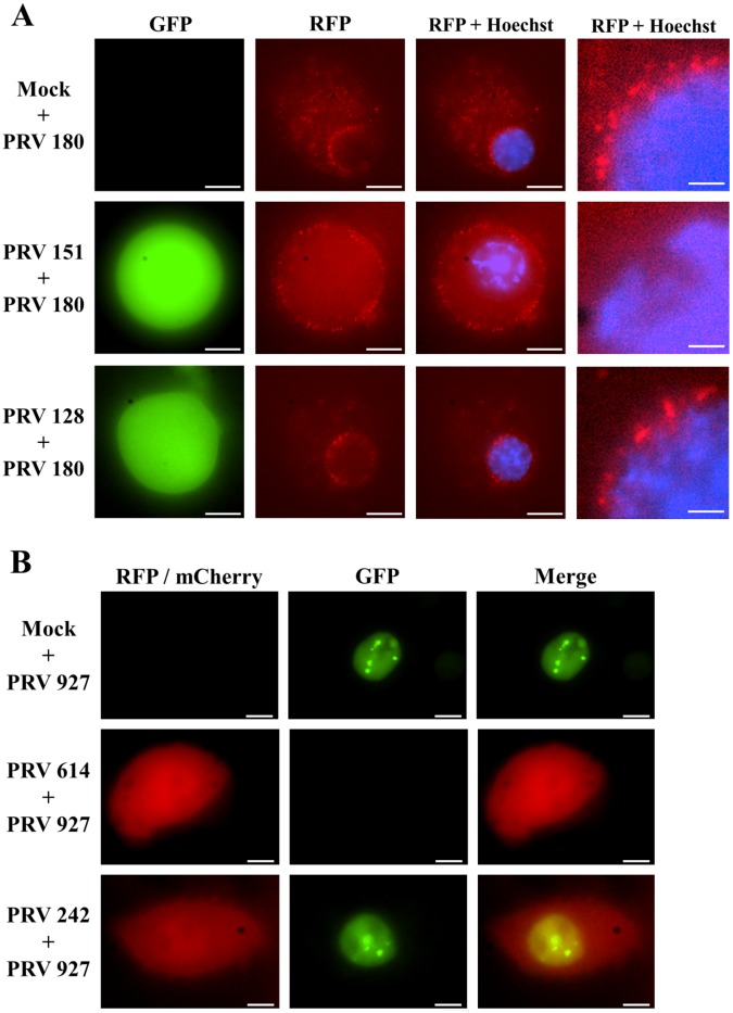
FIG 4 .
IE180-null mutant PRV allows superinfection with a secondary virus. (A) SCG cell bodies were mock infected or infected with PRV 151 or PRV 180 as the primary virus. In all cases, PRV 180 was added 24 h after primary infection and neurons were imaged after 2 h. GFP signal indicates primary infection, RFP signal corresponds to PRV 180 capsids, and blue signal corresponds to Hoechst nuclear staining. The fate of PRV 180 capsids can be observed with more detail in the magnified panel at the right of each row to determine if there was secondary infection, as evidence by capsid entry and transport to the perinuclear region. Scale bar = 10 µm for the first three panels of each row and 2 µm for the magnified inset of each row. (B) PK15 epithelial cells were infected with PRV 614 (red) or PRV 242 (red, IE180-null PRV) as the primary virus (MOI = 10) or mock infected. Eight hours later, cells were infected with PRV 927 (green, ICP8-EGFP PRV) as the secondary virus (MOI = 10). Cells were imaged 6 h after secondary infection. Scale bars = 5 µm.

