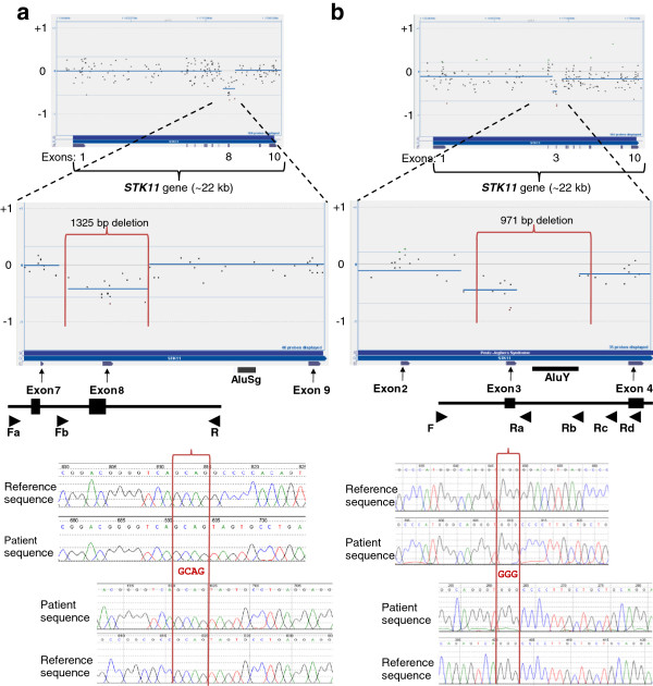
Figure 2.

Single exon deletions in the STK11 gene. Figure 2 shows the data from two independent aCGH analyses with probes targeting the entire STK11 gene, and the zoomed-in view of where the deletion was present. CBS-generated deletion call did not cross the -0.6 threshold in either analysis. The breakpoints of the actual deletion are shown with vertical red lines. Below the CytoSure display are the corresponding exon tracks. Locations of Alu elements in the region of the deletion are marked. At the bottom is an illustration of the breakpoint PCR design, with the location of respective primers shown as arrows. Electropherograms of bidirectionally sequenced deleted alleles are shown with sequencing in the forward direction on top and reverse sequencing below. The four- and three-base pair microhomology at the breakpoints are shown within the two vertical red lines that demarcate the breakpoints. 2a) 1325-bp deletion encompassing exon 8 of the STK11 gene, with electropherogram of sequenced STK11 across deleted exon 8. 2b) 971-bp deletion encompassing exon 3 of the STK11 gene, with electropherogram of sequenced STK11 across deleted exon 3.
