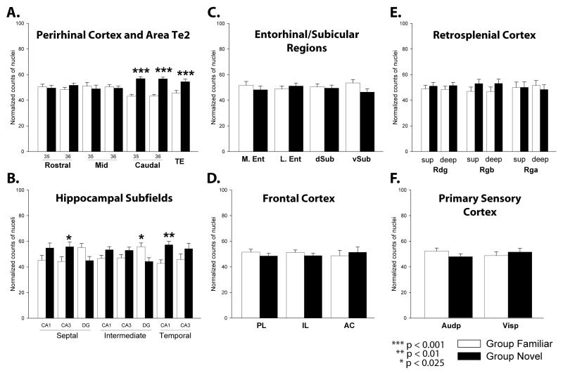
Figure 5.
c-Fos levels following familiar (white) or novel (black) object exposure. A. Perirhinal cortex and Area Te2: areas 35 and 36 of the rostral, mid and caudal perirhinal cortex and area Te2. B. Hippocampal Subfields: dentate gyrus (DG), CA3, CA1, for septal, intermediate and temporal hippocampus. C. Entorhinal/Subicular Regions: lateral entorhinal (lEnt) cortex, medial entorhinal (mEnt) cortex, dorsal subiculum (d Sub) and ventral subiculum (v Sub). D. Frontal Cortex: prelimbic (PL), infralimbic (IL) and anterior cingulate (AC) cortices. E. Retrosplenial Cortex: superficial and deep layers of the dysgranular (Rdg) and granular (Rgb and Rga) retrosplenial cortex. F. Primary Sensory Cortex: primary auditory cortex (Audp), primary visual cortex (Vis). Normalized counts of c-Fos-positive cells are presented as mean ± SEM. Significance of group differences: * p < 0.025; ** p < 0.01; *** p < 0.001.

