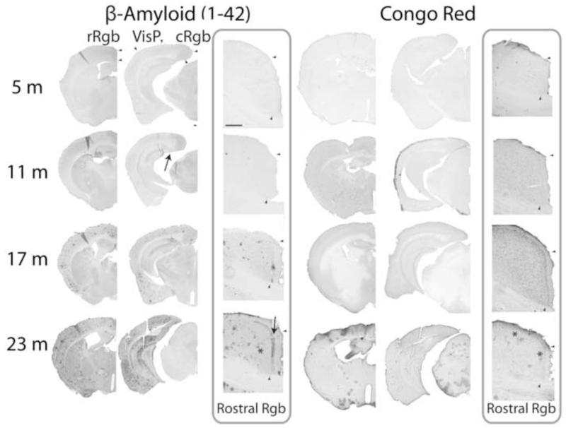
Figure 1. Ageing and amyloid accumulation in Tg2576 mice [β-amyloid (1-42), Congo Red (with Nissl counterstain)].
For each marker, a representative photomontage demonstrates the extent of labeling, while a 5× photomicrograph presents specifically the progression of pathology in the retrosplenial cortex (rostral). Small deposits of β-amyloid (1-42) label can be detected at 11 months sometimes, usually appearing in the subiculum and adjacent forceps major of corpus callosum (arrow). Next, pervasive labeling can be seen at 17 months, including in the retrosplenial cortex, followed with widespread and intense label at 23 months. Notice the distinctive superficial laminar pattern of diffuse extracellular amyloid accumulation seen with increasing age in the retrosplenial cortex (arrow). Congophilic cored plaques appear occasionally in the hippocampal formation at 17 months, extending into the cortex at 23 months, including a few in the retrosplenial cortex (* shows example). Arrowheads indicate the limit of the rostral and caudal granular b retrosplenial cortex (r- and cRgb), including the boundary between superficial and deep laminae, as well as that of the primary visual cortex (VisP). Scale bars = 200 μm.

