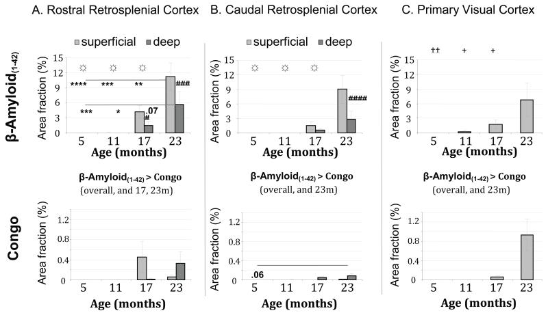
Figure 3. Histograms of amyloid deposition across age groups of transgenic mice.
For rostral retrosplenial (Rgb) cortex, the load of β-Amyloid(1-42) increased with age (‡, between age groups over both markers; *, across age groups for each laminae per marker, **** p < 0.001; *** p < 0.005; ** p < 0.01; * p < 0.05); whereas formation of congophilic material was relatively less abundant (☼, between age groups for single marker). β-Amyloid(1-42) burden was preferentially found in superficial laminae (#, between superficial and deep laminae within age group). Amyloid also accumulated significantly in the caudal portion of the retrosplenial cortex, but it appeared less pronounced than in the rostral portion. In the primary visual cortex, amyloid load was only significant at 23 months, cored congophilic plaques again less abundant than β-Amyloid(1-42)-labeled material. Errors bars represent the standard error of the mean.

