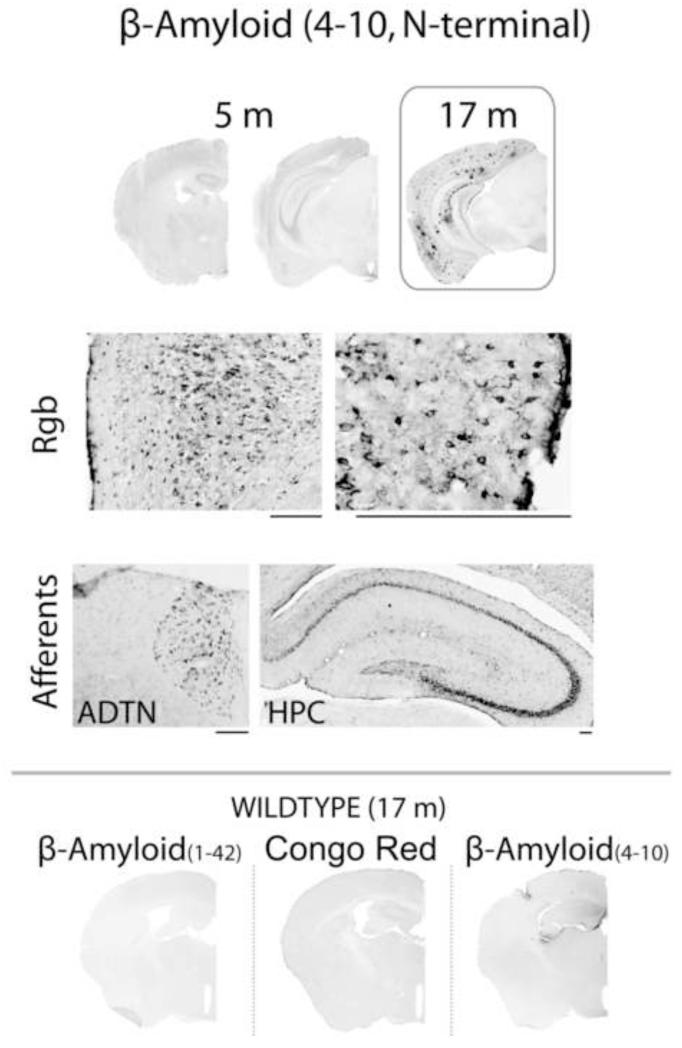
Figure 5. β-amyloid (4-10) pathology in 5-month old APP+ mice.
Marked β-amyloid (4-10) label was observed in the retrosplenial cortex and some of its afferents (top panel, right and inset), months before the appearance of Congo-positive plaques and of material labeled with β-amyloid(1-42) (Fig. 2). The distribution of amyloid accumulation appears to be selectively associated with cells. In contrast, at 17 months the appearance of β-amyloid (4-10) appears equivalent to that of β-amyloid (1-42). Rgb, retrosplenial cortex granular b area; ADTN, anterodorsal thalamic nucleus; HPC, hippocampus. Scale bar = 200 μm.

