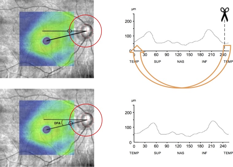
Figure 1.
Calculation of the DFA and compensating for it in RNFL measurements. DFA was calculated on SLO fundus images overlaid with macular thickness maps. Foveal center was found by fitting an ellipse to fovea. To find the disc centroid, the least-squares method was used for fitting an ellipse through a set of eight points identified on the clinical disc margin (left). After calculation of the DFA and rounding it to the nearest whole degree, the TSNIT RNFL measurements were adjusted (right) by the magnitude of the DFA (clockwise for negative angles and counterclockwise for positive angles). More negative DFA values indicate that the fovea is located more inferiorly with respect to the disc. Circles on the left images define the origin of the RNFL TSNIT profile.

