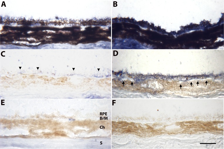
Figure 3.
Immunohistochemistry of ApoB100 of the RPE-choroid in 2-month-old WT mice (A, C, E) and apoB100 (B, D, F) fed a normal chow diet. (A) Unbleached section from a WT mouse from a region showing some immunolabeling for apoB100, mainly in the apical regions of some RPE cells. (B) Unbleached section showing blue immunolabeling for apoB100 throughout RPE cells, which is partially obscured by melanin pigment. The sclera likely stains for apoB100, presumably from systemic deposition, as well as nonspecific staining. (C) Bleached section of a WT mouse showing mild, mosaic apoB100 immunolabeling of the RPE (arrowheads) and BrM. (D) Bleached section from an apoB100 mouse showing strong immunolabeling for apoB100 in the RPE and BrM. The staining appears in every RPE cell in the section. The choriocapillaris endothelium is quiet (arrows). (E) Bleached, nonimmune IgG control of a WT mouse showing mild background staining, especially in the sclera. (F) Bleached, nonimmune IgG control of an apoB100 mouse showing no background staining. Ch, choroid; S, sclera. Scale bar: 20 μm.

