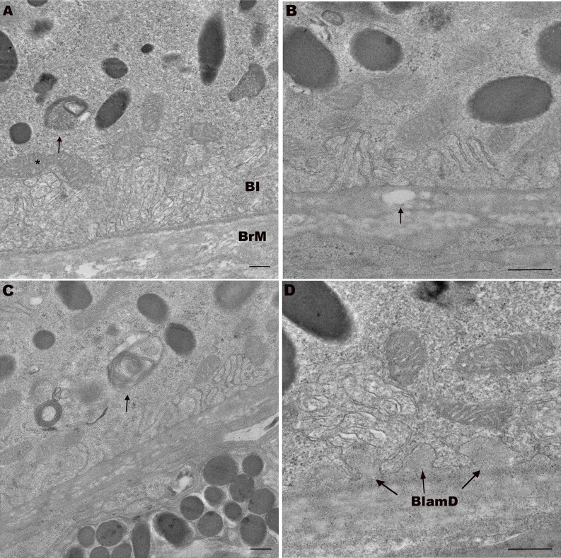
Figure 7.
Transverse electron microscopy of 18-month-old ApoB100 mice shows aging, but no AMD changes. (A) An 18-month-old WT mouse with normal appearing RPE including mitochondria (*) and BI, and BrM. Note undigested photoreceptor outer segment (arrow). (B) An 18-month-old apoB100 mouse with preserved RPE BI. Bruch's membrane has a vacuole that is suggestive of lipid accumulation (arrow). (C) An 18-month-old apoB100 mouse with mild RPE abnormality including partially digested photoreceptor outer segment (arrow) in the basal RPE. Note the preserved basal infoldings. (D) An 18-month-old apoB100 mouse with preserved RPE and thin, homogeneous basal laminar deposits (BlamD, arrows). Scale bar: 300 nm.

