
Figure 3.
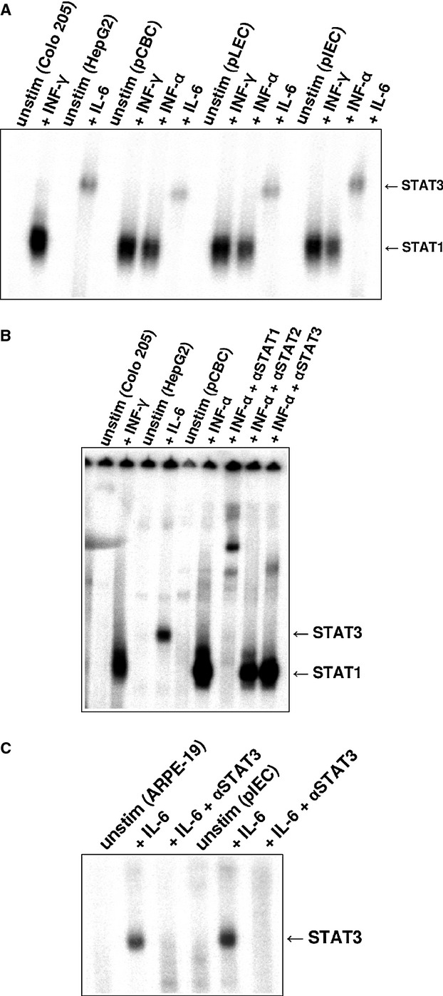
Electromobility shift assay (EMSA) analysis of nuclear extracts from pCBC, pLEC, and pIEC incubated with 32P-labeled M67 probe. (A) Colo 205 cells were left untreated or stimulated with 500 U/ml of human IFN-γ for 20 min STAT1 positive control). HepG2 cells stimulated with 10 ng/ml of human IL-6 were used as STAT3 positive control. pCBC, pLEC, and pIEC cell extracts were either untreated or treated with 10 ng/ml of porcine IFN-γ, with 500 U/ml of human IFN-α or with 10 ng/ml of human IL-6 for 20 min. The position of activated STAT1 and STAT3 proteins is indicated by arrows. (B) EMSA supershift experiment for STAT1 and STAT3 upon stimulation with human IFN-α using M67 probe. pCBC cells were left untreated or stimulated with human IFN-α. IFN-α treated pCBC cells were incubated with antisera specific for STAT1 (IFN-α + αSTAT1), with antisera specific for STAT2 (IFN-α + αSTAT2) or with antisera specific for STAT3 (IFN-α + αSTAT3). The nuclear extracts of human Colo 205 cell line treated with 10 ng/ml of human IFN-γ for 20 min were used as a positive control for the STAT1 and HepG2 cells treated with 10 ng/ml of human IL-6 as a positive control for STAT3. (C) EMSA supershift experiment for STAT3 upon stimulation with IL-6 on M67 probe. pLEC cells were left untreated or stimulated with human IL-6. IL-6-stimulated pLEC cells were incubated with antisera specific for STAT3 (Il-6 + αSTAT3). ARPE-19 cells were used as a positive STAT3 control. The cells were treated the same way as porcine cells: the cells left untreated or were stimulated with human IL-6 for 20 min. ll-6-treated ARPE-19 were incubated with antisera specific for STAT3 (Il-6 + αSTAT3).