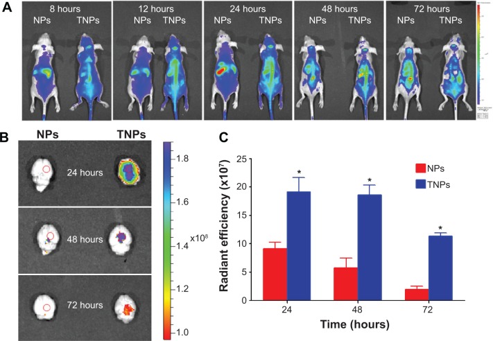
Figure 4.
In vivo imaging. (A) In vivo imaging of brain glioma-bearing nude mice administered DiR-labeled NPs and TNPs at different time points. (B) Ex vivo imaging of the brains at 24, 48, and 72 hours, and (C) the corresponding semiquantitative radiant efficiency at the glioma site.
Note: *P<0.01 between TNP group and NP group.
Abbreviations: NPs, nanoparticles; CREKA, cysteine–arginine–glutamic acid–lysine–alanine; TNPs, CREKA-conjugated NPs; DiR, 1,1′-dioctadecyl-3,3,3′,3′-tetramethylindo tri carbocyanine iodide; PBS, phosphate-buffered saline.

