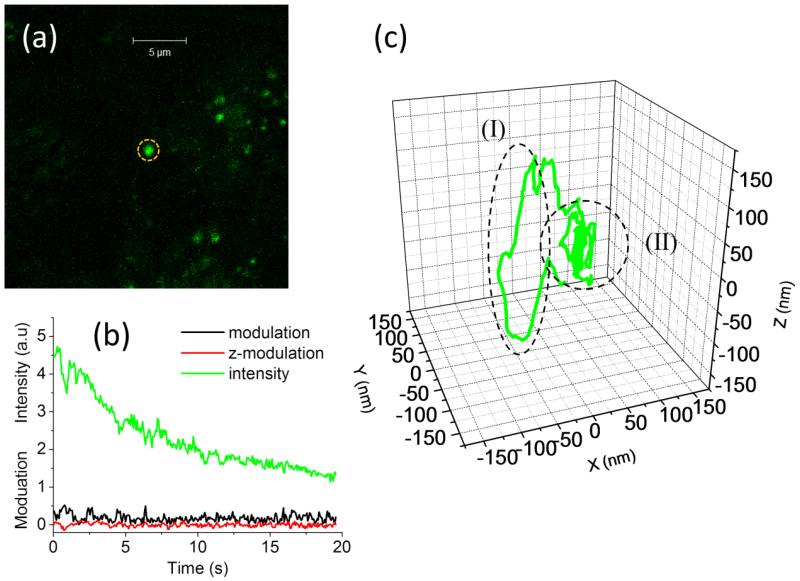
Figure 7.
3D single particle tracking of acidic vesicles in live polarized OK cells. (a) Confocal image of vesicles labeled with pHrodo Green Dextran in OK cells. An isolated vesicle is positioned in center of the field of view (dashed circle). (b) As the tracking algorithm is started, the particle is kept close to the laser beam, as demonstrated by the continuous photobleaching of the intensity and the value of modulation close to zero. (c) In the 3D trajectory we can distinguish a segment of directed motion (dashed oval, I) and a segment of diffusive motion (dashed circle, II).

