
Figure 6.
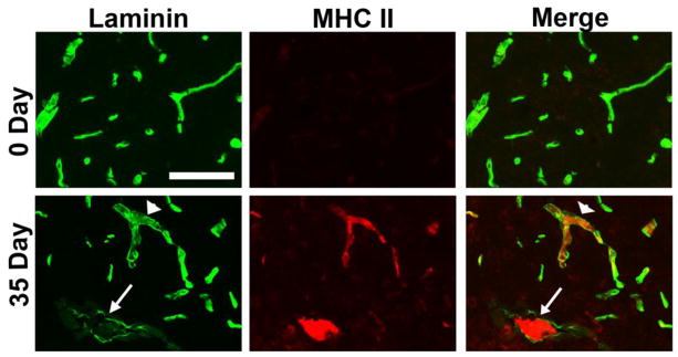
Perivascular cuffing and laminin degradation in post-capillary venules in EAE. A. Dual IF for laminin (Alexa Fluor-488, green) and MHC class II (Cy3, red) was performed on frozen sections of cervical spinal cord taken from 0 and 35 days post-immunization. Scale bar = 100 μm. Note that in chronic EAE (day 35), infiltrating leukocytes were closely associated with post-capillary venules, and that at sites of leukocyte accumulation, vascular laminin staining was greatly reduced (see arrow and arrowhead), suggestive of ongoing proteolysis of basement membrane laminin.