Figure 1.
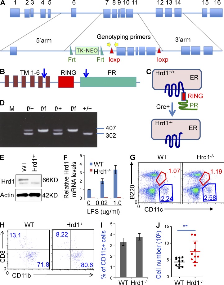
Targeted disruption of the Hrd1 gene in DCs. (A) Structures of the Hrd1 WT and targeted alleles. Exons and the neomycin phosphotransferase gene (Neo) driven by the thymidine kinase (TK) promoter are shown. The TK-NEO cassette is flanked by 2 FRT sites and exons 7–11 are flanked by 2 LoxP sites. (B and C) Domain structure of Hrd1 protein. The ER membrane-anchoring protein Hrd1 carries 6 transmembrane (TM) domains, one RING finger domain, and a C terminus proline-rich domain. The deletion of floxed Hrd1 gene by Cre recombinase destroys Hrd1 protein expression. (D) Genotyping of Hrd1-floxed mice. Tail snips from a litter of Hrd1flox/wt X Hrd1flox/wt offspring were collected for DNA extraction and PCR analysis. The 302-bp PCR product is the WT allele and the 407-bp product is the mutant allele. (E and F) BM cells were isolated from WT and Hrd1 conditional KO (Hrd1−/−) mice and cultured under DC differentiation conditions. (E) Hrd1 protein expression was analyzed by immunoblotting (top) using β-actin as a loading control (bottom). (F) Hrd1 mRNA levels were determined by real-time quantitative RT-PCR. Hrd1 levels in WT DCs increased with LPS treatment. (G) Cell surface expression of B220 and CD11c in total splenocytes from WT and Hrd1−/− mice was analyzed by flow cytometry. (H) CD11c+B220− cells were gated and the expression of CD8 and CD11b was analyzed. (I and J) The mean percentages (I) and absolute numbers (J) of CD11c+ cells in the spleens of 10 pairs of WT and Hrd1−/− mice are shown (n = 10).

