Figure 5.
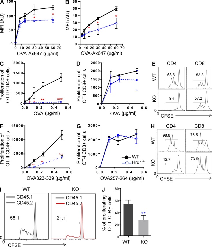
Hrd1 deficiency impairs DC-mediated CD4+ T cell priming. (A and B) BMDCs (A) and sorted CD11c+ cells (B) from the spleen were cultured with Ax647-OVA at the indicated concentrations for 1 h and washed. Fluorescence intensity was analyzed by flow cytometry and data are reported as mean ± SD from five independent experiments. (C–E) BMDCs were stimulated with LPS in the presence of OVA protein overnight, washed, and co-cultured with either CD4+ OT-II T cells or CD8+ OT-II T cells for 3 d. T cell proliferation was analyzed by 3H-thymidine incorporation (C and D) or CFSE dilution (E). (F–H) BMDCs were stimulated with LPS in the presence of either OVA323-339 (F and H [left]) or OVA257-264 (G and H [right]) peptides overnight, washed, and co-cultured with either CD4+ OT-II T cells or CD8+ OT-II T cells for 3 d. T cell proliferation was analyzed by 3H-thymidine incorporation (F and G) or CFSE dilution (H). (I and J) In vivo OVA-specific CD4+ T cell priming was analyzed as described in Materials and methods. (I and J) Representative images (I) and data reported as mean ± SD (J) from five pairs of WT and Hrd1−/− recipients are shown (n = 5). Student’s t test was used for statistical analysis. *, P < 0.05; **, P < 0.01; ***, P < 0.0001.

