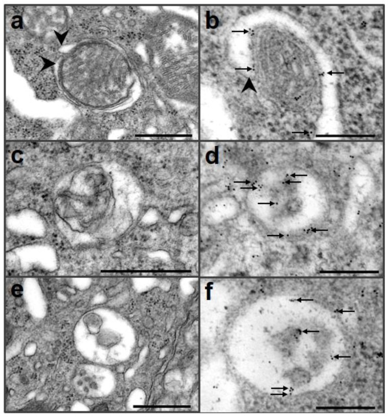
Figure 2.
Immunogold electron microscopic analysis of autophagy in Purkinje cells of Ngsk mice using an anti-LC3 antibody. Autophagosomal membranes (arrowheads) began to form vacuoles that contained mitochondria (a, b), and gold particles (arrows) were located in isolated membranes (b). Autophagolysosome showed that single membrane surrounding mitochondrion (c). Immunogold particles (arrows) were located in single membrane and residues (d). Autophagolysosomes were bounded by a single membrane and contained residues (e), and gold particles (arrows) were detected in single membrane and residues (f). Scale bar = 500 nm (a, c, e), 200 nm (b, d, f).

