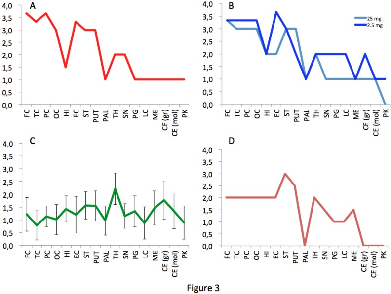
Figure 3.
Spongiosis profiles in infected primates. Lesional profiles (based on spongiosis) in primates exposed to cattle-adapted TME (A) L-BSE (B), c-BSE (C) and raccoon TME (D) were defined according to the scoring and areas described by Parchi et al. [27]. Spongiosis profile of c-BSE primates is depicted as the mean among 5 primates exposed to c-BSE. Frontal Cortex (FC) ,Temporal Cortex (TC), Parietal Cortex (PC), Occipital Cortex (OC), Hippocampus (HI), Entorhinal Cortex (EC),Striatum (ST), Putamen (PUT), Pallidum (PAL), Thalamus (TH), Substantia Nigra (SN), Periventricular Gray (PG), Locus coeruleus (LC), Medulla (ME), Cerebellum (granules) (CB), Cerebellum (molecular layer) (CB), Purkinje cells (PK).

