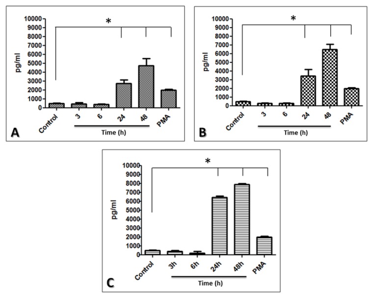
Figure 6.
Production of IL-6 by limbo-corneal fibroblasts infected with mycobacteria. The limbo-corneal fibroblasts were infected with MTB, MAB, and MSM for 3 h, and the kinetics of the infection were followed for 6 h, 24 h, and 48 h. The supernatants were recovered at each post-infection time. The production of IL-6 in the supernatants was quantified using CBA. The levels of IL-6 induced by the infection are presented as pg/ml; the PMA-stimulated production of IL-6 is included in the graph. The results are expressed as the mean ± SD for 2 independent experiments (*P< 0.05). A) IL-6 induced by MTB, B) IL-6 induced by MAB, and C) IL-6 induced by MSM.

