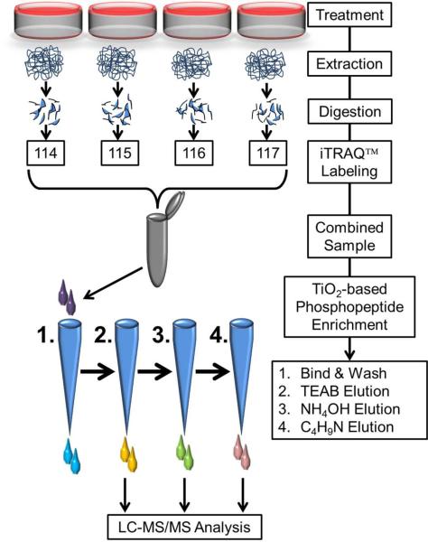
Figure 1.
Schematic workflow of sample preparation for quantitative phosphoproteomics. Cultures were treated with or without PMI 5011 in the presence or absence of insulin and proteins were extracted and digested with trypsin. After iTRAQ™ labeling, the combined peptide samples were enriched for phosphopeptides. In order to avoid overloading of the TiO2 columns, the iTRAQ™ sample was split in four equal parts and loaded on four microcolumns. All four columns were first washed and then eluted sequentially with triethyl ammonium bicarbonate, ammonium hydroxide and pyrolidine solutions in acetonitrile. Same solvent elutions from the four columns were combined, dried under vacuum and resuspended in 97% H2O, 3% acetonitrile and 0.1% formic acid prior to mass spectrometry analysis.

