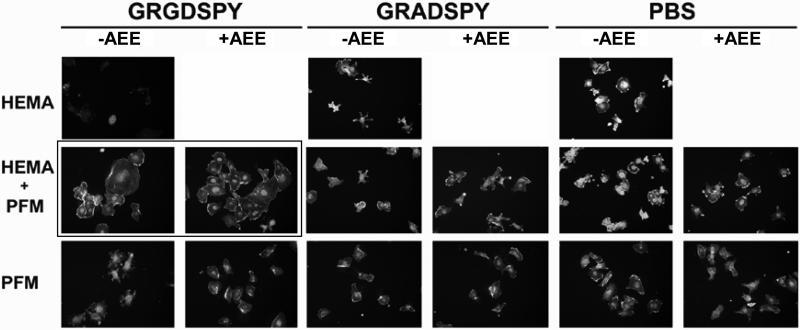
Figure 11. HUVEC morphology at 2 h of incubation.
Cells were cultured on three different substrates: the HEMA-based hydrogel (first raw), the hydrogel with a PFM layer (second raw) and the PFM polymer (third raw). The films were functionalized with GRGDSPY (first and second column) and GRADSPY (third and forth column) and, as controls, other samples were immersed in PBS under the same conditions (fifth and sixth column). ‘+AEE’ and ‘-AEE’ indicate whether or not the samples were treated with the passivating amine.

