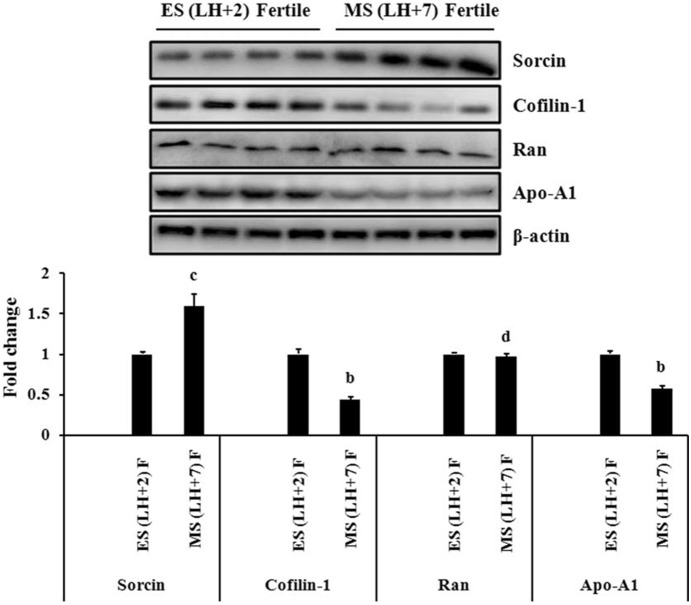
Figure 4. Immunoblot analysis of Sorcin, Cofilin-1, Apolipoprotein-A1 (Apo-A1) and Ran GTP-binding nuclear protein (Ran) in fertile women.
Expression of Sorcin, Cofilin-1, Apo-A1 and Ran were checked in early-secretory (LH+2) and mid-secretory (LH+7) phase endometrium of fertile women. Representative images (upper panel) of immunoblot of Sorcin, Cofilin-1, Apo-A1 and Ran have been shown. β-actin served as a loading control for normalization. Quantification of band intensity (lower panel) was performed by densitometric analysis by using Quantity One software (v. 4.5.1) and a Gel Doc imaging system (Bio-Rad). p-values are (b) p<0.01, (c) p<0.05 and (d) p>0.05 versus early-secretory phase (LH+2). Values are expressed as mean± SE; n = 4, ES = early-secretory phase, MS = mid-secretory phase, LH+2 = 2 days after luteinizing hormone surge, LH+7 = 7 days after luteinizing hormone surge.

