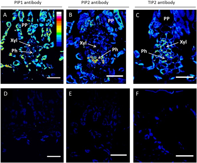
Figure 7. Immunolocalization of AQP proteins in leaves of P. trichocarpa saplings.
Confocal laser scanning micrographs showing the localization of PIP1, PIP2, TIP2 proteins in minor veins of leaf transverse sections (A, B, C respectively). Controls with no primary antibody indicate minimal background fluorescence (D, E, F respectively). Images were taken at an identical setting and were color-coded with an intensity look-up-table (LUT; displayed in A), in which black was used to encode background, and blue, green, yellow, red and white to encode increasing signal intensities. Ph, phloem; PP, palisade parenchyma; Xyl, xylem. Scale bars = 20 µm.

