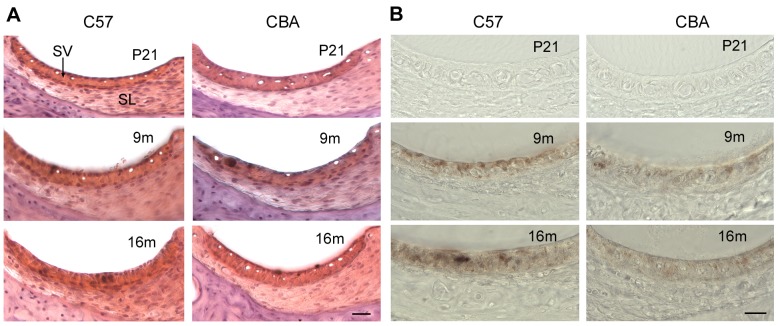
Figure 2. Cross sections of the LW at three different times points during age.
Sections were obtained from cochlear middle turns at P21, 9 m, and 16 m. A. Cross sections stained with Hematoxylin and Eosin. Scale bars: 20 µm. B. Cross sections without any staining. Hyperpigmentation and reduction in the number of capillaries are apparent in the SV at 16 m.

