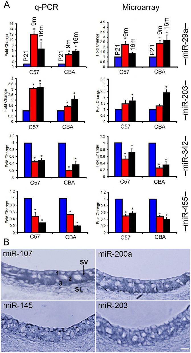
Figure 6. miRNA expression detected by q-PCR and in situ hybridization.
A: Comparison of changes in miRNA expression detected by q-PCR versus microarray analyses for four miRNAs in the SV of C57 and CBA mice. Asterisks indicate statistically significant differences (p<0.05) compared to P21. Each q-PCR plot represents means from three repeats. B: Expression of four miRNAs in the LW of C57 mice using in situ hybridization technique. SV: Stria vascularis. SL: Spiral ligament. 1, 2, and 3 mark the three cell types in the SV (marginal, intermediate, and basal cells), respectively. Bar: 20 µm.

