
Figure 3. MR-angiographic and duplex sonographic images of the great saphenous vein.
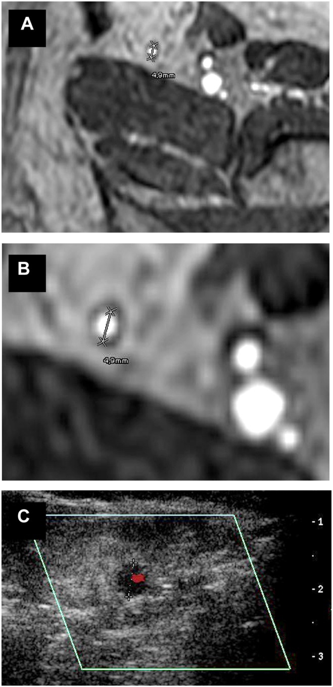
Magnetic resonance imaging (BPCA-MRA) and color-coded duplex sonography in the proximal level of the left GSV of a 63 year old female patient who suffered from PAOD stage III and was referred to the radiological department for assessment of the arterial status prior to a proposed bypass surgery. (a, b) Axial multiplanar reformat of contrast-enhanced T1-weighted gradient-echo images during the steady-state. (c) Axial color-coded duplex sonography.