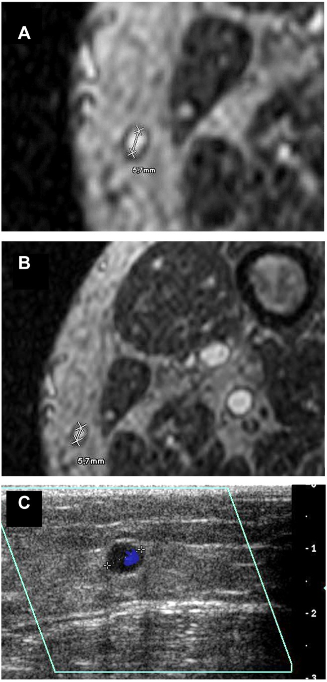
Figure 4. MR-angiographic and duplex sonographic images of the great saphenous vein.

Magnetic resonance imaging (BPCA-MRA) and color-coded duplex sonography in the distal level of the left GSV in a 69 year old male patient with PAOD stage IV. (a, b) Axial multiplanar reformat of contrast-enhanced T1-weighted gradient-echo images during the steady-state. (c) Axial color-coded duplex sonography.
