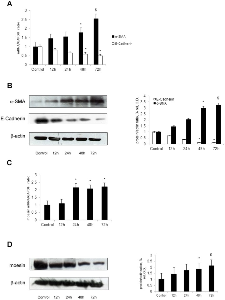
Figure 1. TGF-β1 up-regulates moesin and α-SMA, down-regulates E-Cadherin in HK-2 cells.
HK-2 cells were maintained in the absence or presence of TGF-β1 (5 ng/ml) for various hours. The cells treated with TGF-β1 presented with up-regulated expression of α-SMA and down-regulated expression of E-Cadherin by real-time PCR (A) and western blot (B) in comparison with control. TGF-β1 also upregulated moesin expression in HK-2 cells for indicated time period. The expression of moesin was determined by real-time PCR (C) and western blot (D). β-actin was used to verify equivalent loading. Densitometrical analysis and real-time PCR results shown were results from three independent cell preparations. Western blot showed the results from one of three independent preparations. * p<0.05 versus Control; $ p<0.01 versus Control.

