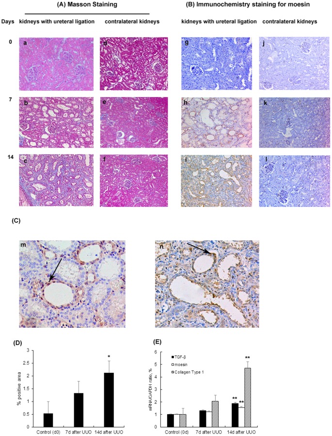
Figure 2. Expression of moesin in rat model of UUO.
Kidney histology showed the histological injury of the rats. The rats had left kidneys ureteral ligation and right kidneys were set as control. (A) On masson stained sections (x100), the kidneys were normal at day 0 (a). One week after the left ureteral ligation, rats developed minor tubulointerestital injury which included minor tubular atrophy and mild collagen deposition (b). Two weeks after the ligation, severe tubulointerestital injury was found, including severe tubular atrophy and large amount of collagen deposition (c). (B) Immunohistochemical staining (x100) in rats' kidneys showed that there was barely moesin detected in the kidneys at day 0. There was increase of moesin staining in the tubulointerestitium at day 7 and day 14 which were in accordance with tubulointerestital injury. (C) Immunohistochemical staining (x200) also showed that the expressions of moesin in the kidneys with ureteral ligation were mainly localized in renal tubular epithelia cells. Arrows indicated moesin positively stained tubular epithelia cells at day 7(m) and day 14(n) after UUO. (D) Quantification of tubulointerestital fibrosis by using ImageJ software for Masson positively stained areas. (E) TGF-β, collagen type I and moesin mRNA expression in rat kidneys with ureteral ligation by real time PCR. * p<0.05 versus control group (day 0); ** p<0.01 versus control group (day 0).

