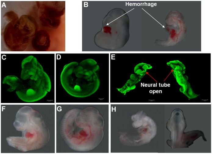
Figure 2. Disruption of pdia3 gene resulted in malformation during embryogenesis.
The partially absorbed embryos were observed at E12.5 (A). E10.5 embryos were dissected and observed under stereomicroscopy (B, F–H) and confocal microscopy after acridine orange staining (C–E). The wild type embryos (C, F) and heterozygous embryos (D, G) were similar in size and larger than homozygous embryos. One homozygous embryo displayed hemorrhage around the heart area (B) and another one showed open neural tube (E, H). Scale bar = 1000 µm.

