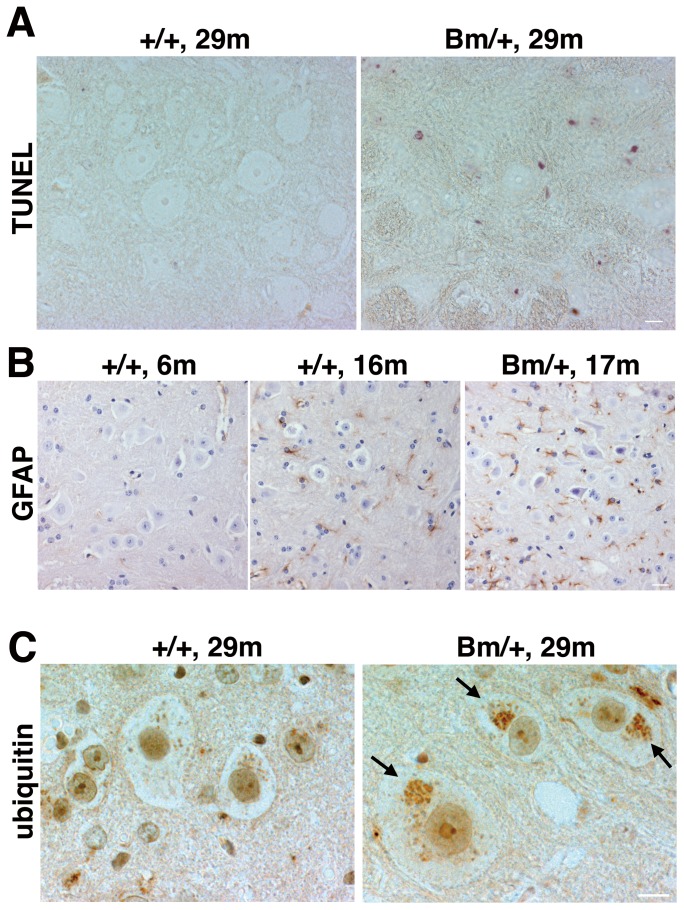
Figure 3. Some motoneurons in the spinal cord revealed a degeneration accompanied by accumulations of ubiquitinated proteins.
(A) TUNEL staining revealed some apoptotic cells at the anterior horn in the spinal cord of a 29 month-old mutant BiP mouse (Bm/+, 29 m). Scale bars, 10 um. (B) The immunoreactivity with an anti-GFAP antibody at the anterior horn is increased in a 17 month-old mutant spinal cord (Bm/+, 17 m). Scale bars, 20 um. GFAP positive cells are counted (five fields in each mouse, GFAP positive cells/the number of nucleus). +/+, 16 m; 33/199, 35/174, 34/192, 40/181, 42/207, Bm/+, 17 m; 42/132, 50/140, 59/147, 39/154, 58/143 +/+, 6 m; 5/143, 4/131, 4/141, 0/95, 0/107. The ratio of GFAP positive cells is significantly higher in the mutant BiP spinal cord (Bm/+, 17 m) compared to that in the wild type (+/+, 16 m) by t test (p value is 0.0009). (C) The aggregations were stained by an anti-ubiquitin antibody in the large cells at the anterior horn of the 29 month-old mutant spinal cord (Bm/+, 29 m, arrowheads). Scale bars, 10 um.

