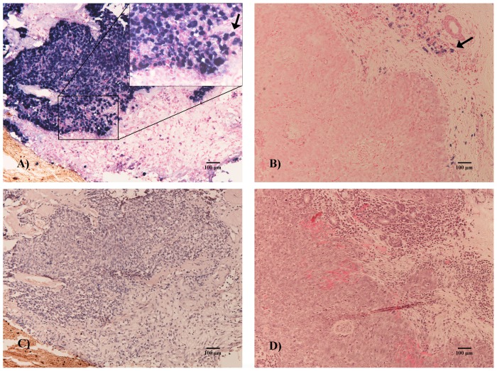
Figure 2. EBV encoded RNA in in situ hybridization.
Micrograph A: most of the neoplasm cells were positive (nasopharyngeal carcinoma, ×100, bar = 100 µm), as indicated by the arrow at high magnification (×200). Micrograph B: although some positive lymphocytes are apparent (arrow), no neoplasm cells were positive. Micrographs C and D: Sections stained with hematoxylin and eosin (HE).

