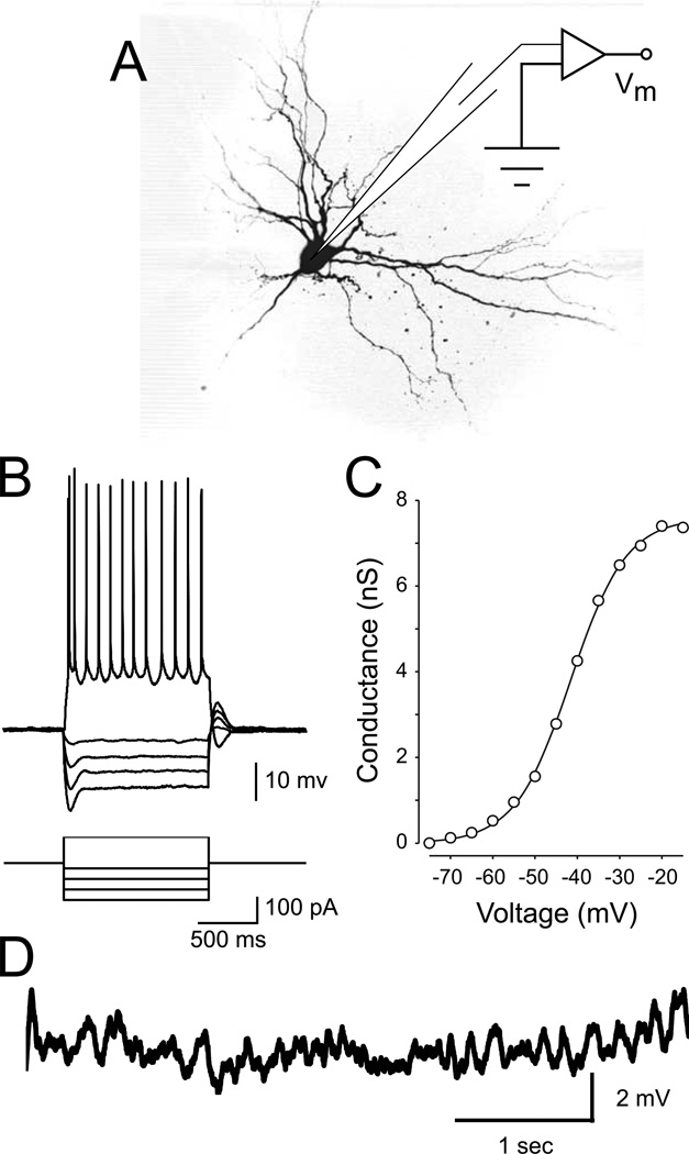
Figure 1. Stellate neuron morphology and electrophysiology.
A Schematic of the recording setup and morphology of a representative stellate neuron. B Stellate neurons display a pronounced inward rectification (sag) in response to hyperpolarizing current steps. In response to a step of depolarizing current, stellate neurons respond with a short burst of action potentials followed by tonic spiking. C Average activation curve of persistent sodium conductance (GNaP) across a population of stellate cells (modified from Burton et al., 2008). D Spontaneous subthreshold oscillations appear as the neuron is depolarized to a just below spike threshold.

