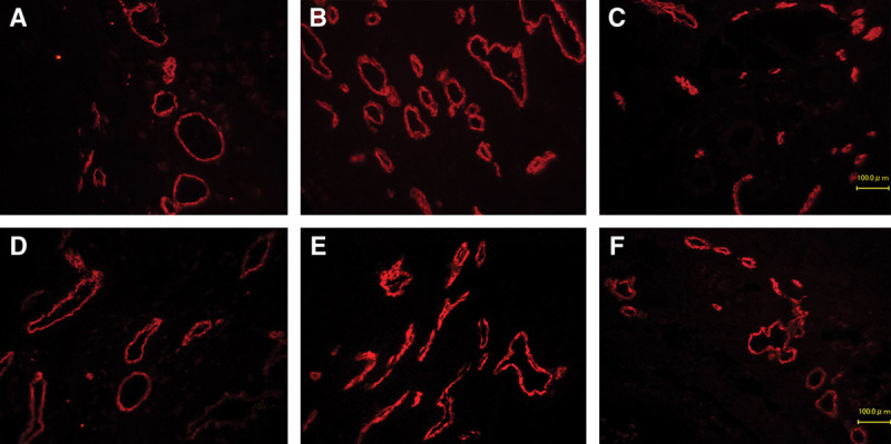
Fig. 9.

Immunohistochemical fluorescence staining for LYVE1. The numbers of visible lymphatic vessels (red) gradually increased over time. Lymphatic vessels are mainly located in subcutaneous tissue above fascia. A, bFGF-POD 7; B, bFGF-POD 14; C, bFGF-POD 28; D, Control-POD 7; E, Control-POD 14; F, Control-POD 28 (×100).
