
Figure 4.
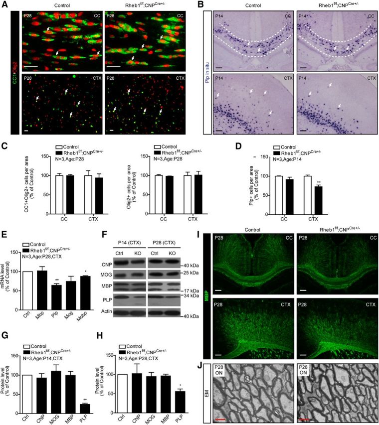
Loss of Rheb1 in Rheb1f/f; CNPCre+/− mice does not affect differentiation of OPCs but does affect certain components of myelin. A, Immunofluorescence of CC1+/Olig2+ cells (arrows) in the corpus callosum (CC) and cortex (CTX) of P28 control and Rheb1f/f;CNPCre+/− brains. Scale bar, 40 μm. B, In situ hybridization showing Plp+ cells (indicated by arrows) in brain of control (Rheb1f/+; CNPCre+/− or Rheb1+/+; CNPCre+/−) and Rheb1f/f;CNPCre+/− mice in the corpus callosum outlined by dashed lines and cortex at P14. Scale bar, 100 μm. C, Quantification of OL lineage cells (Olig2+) and differentiated OLs (CC1+/Olig2+) in the corpus callosum and cortex of P28 animals (mean ± SEM, n = 3). D, Quantification of Plp+ cells in the corpus callosum and cortex of Rheb1f/f;CNPCre+/− mice at P14 (mean ± SEM, n = 3,**p = 0.0061). E, mRNA expression of myelin genes in the cortex at P28 control and Rheb1f/f;CNPCre+/− mice (Mbp, p = 0.8836; Plp, **p = 0.0073; Mog, p = 0.2008; Mobp,*p = 0.0121; mean ± SEM, n = 3). F–H, Western blot analysis of myelin protein expression in the cortex of control and Rheb1f/f;CNPCre+/− mice at P14 (left panel of F, G) and P28 (right panel of F, H). All quantifications are expressed as percentage control ± SEM; **p = 0.0015, *p = 0.0201, n = 3. I, MBP staining reveals no dramatic reduction of MBP-positive fibers in the CC and CTX of Rheb1f/f;CNPCre+/− mice at P28. Scale bars: 100 μm. J, EMs from the optic nerve (ON) of control and Rheb1f/f;CNPCre+/− mice at P28. Scale bar, 500 nm.