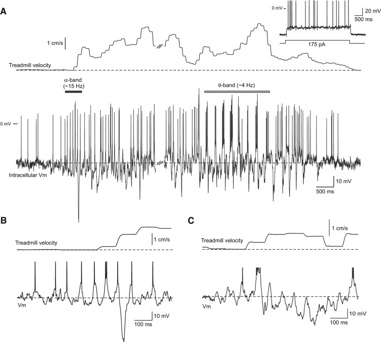
Figure 1.
Intracellular response recorded during a spontaneous movement epoch. (A) Simultaneous recording of intracellular membrane potential in a dentate gyrus neuron (bottom trace) and spherical treadmill velocity (combined forward/backward and rotational axes) during a spontaneous movement epoch (18-sec duration; traces interrupted at double slash mark). Intracellular recording example shows both α-band membrane potential modulation near movement onset (horizontal black bar) and θ-band modulation (gray bar) later during the epoch. Inset shows response to a depolarizing current step in the same neuron. (B) Enlargement of the α-band membrane potential modulation from the example epoch shown in A. (C) Similar α-band membrane potential modulation recorded during movement onset in a different dentate gyrus neuron. Action potentials truncated in B and C.

