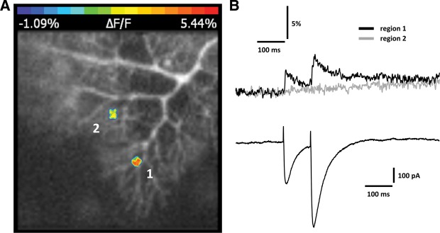
Figure 2.

PF stimulation elicits spatially restricted calcium transients. (A) Original image showing two regions of interest (ROI) indicating spatial restriction of calcium transient hotspots (false-color coding). Calcium transients are reported as normalized fluorescence changes (ΔF/F). (B) PF-EPSCs (bottom) and associated calcium transients (top) recorded from regions 1 and 2 as shown in A. Fluorescence measurements were performed using an ultra-high-speed CCD camera (NeuroCCD-SMQ; RedShirtImaging) and the fluorescent calcium indicator Oregon Green BAPTA-2 (200 μM).
