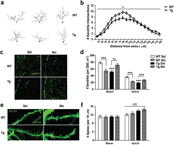
Figure 1.

CA1 pyramidal neurons structure in mice overexpressing the CHRNA5/A3/B4 region and upon chronic nicotine treatment. (a) Representative reconstructions of the apical dendritic tree of Lucifer Yellow transfected wild type (WT, upper panel) and TgCHRNA5/A3/B4 neurons (Tg, lower panel), Scale bar 25 μm. (b) Sholl analysis indicated that transgenic neurons showed reduced number of dendritic intersections compared to WT, particularly at 50 to 150 μm from the cell soma (n = 5–10 cells/animal; 4–5 animals/group from ≥ 3 experiments). (c) Representative confocal images of the CA1 region in Thy1-Yellow Fluorescent Protein (YFP) - WT and Tg mice that received either saline (Sal) or nicotine (Nic, 3.25 mg/Kg/d) for 7 d, Scale bar 25 μm. (d) Quantification of the number of apical and basal dendritic structures in 50 × 50 μm2 area revealed significant reductions in Sal-treated Thy1-YFP-Tg mice, compared to Sal-treated Thy1-YFP-WT mice. Chronic administration of nicotine restored the dendritic deficit in Thy1-YFP-Tg mice but instead the same treatment reduced the number of dendritic structures in Thy1-YFP-WT mice (n = ≥100 images animal; 4–5 animals/group from ≥ 3 experiments). (e) Representative photomicrograph illustrating dendritic spines on apical dendrites in CA1 pyramidal neurons from saline and nicotine treated Thy1-YFP- WT and Tg mice, Scale bar 10 μm. (f) Quantification of spines per 10 μm of dendrite length indicated that apical but not basal dendrites from saline- and nicotine-treated Thy1-YFP-Tg mice presented increased spines as compared to Thy1-YFP-WT (n = 5–10 cells/animal; 4–5 animals/group from ≥ 3 experiments). *p ≤ 0.05, **p ≤ 0.01, ***p ≤ 0.001; Two-way ANOVA genotype effect σσ p ≤ 0.01.
