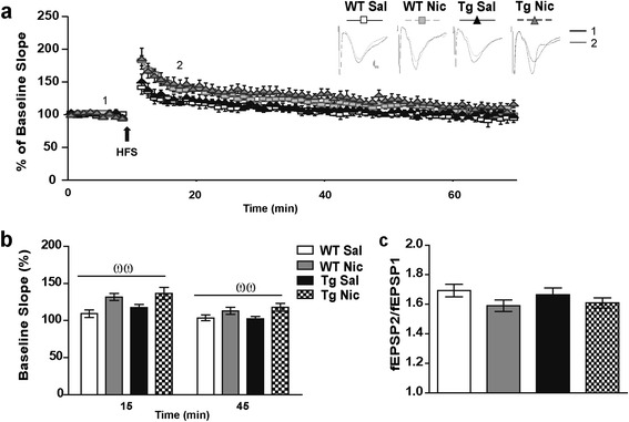
Figure 3.

Electrically induced synaptic plasticity of Shaffer collateral-CA1 pathway. (a) High frequency stimulation (HFS; 100Hz, 1s) induced synaptic potentiation at Schaffer Collateral (SC)-CA1 synapses similarly in slices from saline (Sal)-treated wild type (WT) and TgCHRNA5/A3/B4 (Tg) mice. Chronic nicotine (Nic, 3.25 mg/Kg/d) administration for 7 d enhanced synaptic potentiation both in WT and Tg mice. Upper panel: representative field excitatory postsynaptic potentials (fEPSPs) in response to one stimulation in the CA1 stratum radiatum before (1) and 15 min after (2) applying HFS (arrow), Scale bar 0.5 mV and 1 ms. Lower panel: slopes of the fEPSPs normalized to those obtained before applying HFS (%). (b) Mean values of fEPSPs potentiation at 15 and 45 min after HFS. (c) The paired pulse ratio (fEPSP2/fEPSP1) with an inter-pulse interval of 50 ms (response average of 5 paired pulses; time interval 1 s) was similar among slices from the different experimental conditions (n = 3–5 recordings/animal; 4 – 5 animals/group from ≥ 3 experiments). Two-way ANOVA treatment effect ωω p ≤ 0.01.
