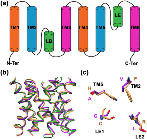
Figure 1.

Topology, structure and selectivity filter of MIP channels. (a) Topology diagram of a MIP channel exhibiting six transmembrane segments (TM1 to TM6) and the two half-helices formed by the functionally important loops LB and LE. (b) Superposition of three MIP channel structures. Water-transporting AQP1 (brown; PDB ID: 1J4N) and glycerol-specific GlpF (green; PDB ID: 1FX8) are superposed on a modeled fungal MIP structure (purple). Only the helical backbone of transmembrane segments and the loop LB and LE are shown for clarity. (c) Superposition of residues forming the aromatic/arginine selectivity filter from the water-transporting AQP1 (brown) and a model from a representative example of SIP-like fungal MIP channel (purple). Amino acids in one letter codes are labeled in the respective color of each structure. The contributing transmembrane segments (TM2 or TM5) and the loop positions (LE1 or LE2) are also indicated.
