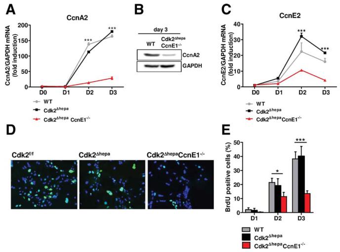
Fig. 2.
Combined depletion of Cdk2 and CcnE1 results in strongly reduced CcnA2 expression and impaired DNA synthesis. Primary mouse hepatocytes from Cdk2f/f (WT), Cdk2Δhepa, and Cdk2ΔhepaCcnE1−/− mice were isolated and cultured for up to 3 days in the presence of EGF and insulin. (A) qPCR analysis of CcnA2 gene expression. (B) Down-regulation of CcnA2 in Cdk2ΔhepaCcnE1−/− mice at day 3 was confirmed by western blotting. (C) qPCR analysis of CcnE2 gene expression. (D) Determination of BrdU incorporation (green) in primary hepatocytes after 3 days in culture on coverslips. Total nuclei are counterstained with 4′, 6-diamidino-2-phenylindole (blue). (E) Quantification of BrdU-positive hepatocytes. *P < 0.05; ***P < 0.001.

