Figure 6.

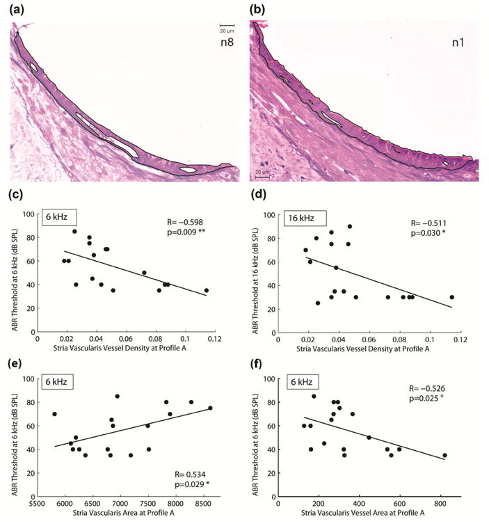
Percentage of IHC (a) and OHC (b) present, spiral ganglion neuron (SGN) density (c), and stria vascular (SV) density (d) in Profile A (electrode region). Only stria vascular density was associated with the high-frequency threshold shifts as shown in Figure 7.
