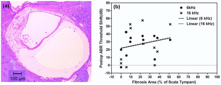
Figure 8.

Example images of cochleae with and without ossification at the cochleostomy site. (a) n5 in the CAES group, showing ossification at cochleostomy site (black arrow). (b) n8 in the NS group without ossification. Note that the electrode array can be seen clearly in this cochlea.
