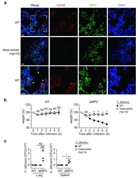
Figure 3. Selective NETosis is critical for clearance of hyphae in vivo.
(a) NET release in the lungs of WT (C57BL/6) mice infected intratracheally with 1×10⁵ c.f.u. WT C. albicans or a hgc1Δ yeast-locked mutant and assessed 24 h post infection by immunofluorescence microscopy for citrullinated histone H3 (Cit-H3, red), MPO (green) and DNA (DAPI, blue). White arrows depict areas of NET release. Lower panel: magnification detail in upper panel. Scale bars = 20 μm. (b) Weight of WT (C57BL/6) (n=6) and MPO deficient (n=5) mice infected with 1×10⁴ c.f.u. WT C. albicans or a hgc1Δ yeast-locked mutant. Weight normalized to staring weight at d0. Statistics by two-way ANOVA, followed by Sidak’s multiple comparison post test: NS p>0.5, ** p<0.0001. (c) C. albicans load in the lung and spleen 6 days post infection (n=3). Statistics by two-way ANOVA, followed by Tukey’s multiple comparison post test: NS p>0.5, * p<0.01. Data are representative of two independent experiments.

