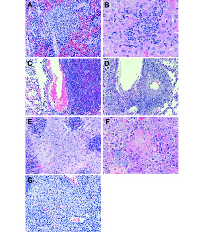
Figure 1.
(A) Spleen from a BALB/c mouse (no. 060231) d after aerosol infection with Francisella tularensis type A. There is a focus of pyogranulomatous inflammation (equal numbers of macrophages and neutrophils) present in the red pulp. Original magnification, 40 ×. (B) Liver from a BALB/c mouse (no. 060231) 3 d after aerosol infection with Francisella tularensis type A. There is a focus of pyogranulomatous inflammation (equal numbers of macrophages and neutrophils) with a few degenerated hepatocytes, evidenced by their bright pink cytoplasm. Original magnification, 60×. (C) Lung from a C57BL/6 mouse (no. 060239) 3 d after aerosol infection with Francisella tularensis type A. There is an area of pneumonia to the right of a blood-filled (artifact) bronchiole, and it is evident that inflammatory cells have invaded the wall of the bronchiole. There is relatively normal lung to the left of the bronchiole. Original magnification, 20×. (D) Lung from a C57BL/6 mouse (no. 060247) 5 d after aerosol infection with Francisella tularensis type A. This field is similar to that in panel C. Note the necrosis of most of the inflammatory cells at this time point. Original magnification, 20×. (E) Spleen from a C57BL/6 mouse (no. 060248) 5 d after aerosol infection with Francisella tularensis type A. Most of the red pulp of this spleen is filled with necrotic inflammatory cells and fibrin (pink material). The blue cellular clusters are remnants of white pulp that has been markedly depleted. Original magnification, 40 ×. (F) Liver from a C57BL/6 mouse (no. 060248) 5 d after aerosol infection with Francisella tularensis type A. Centrally is a group of necrotic hepatocytes with no evidence of attending inflammatory cells. Surrounding this focus, note enlarged cells with blue granular cytoplasm. These are Kupffer cells (and possibly some hepatocytes) that contain Francisella. Original magnification, 40×. (G) Submandibular lymph node from a C57BL/6 mouse (no. 060246) 3 d after aerosol infection with Francisella tularensis type A. There are coalescing foci of inflammation in this lymph node and the inflammation is comprised mostly of macrophages. Many of the macrophages are necrotic as evidenced by the blue nuclear debris. Original magnification, 40×.

