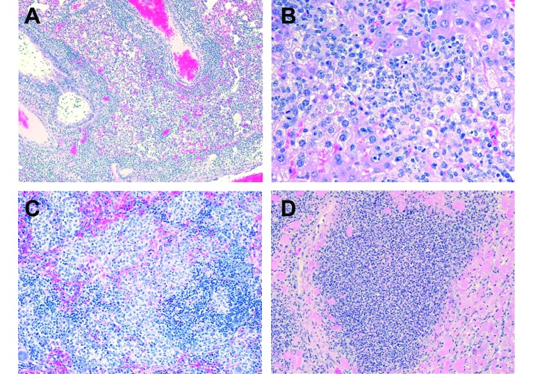
Figure 2.
A. Lung from a BALB/c mouse 12 d following aerosol infection with Francisella tularensis type B. Note the cuffs of lymphoid cells surrounding the blood vessel (near top center) and the bronchiole (to the left of it). Alveoli contain variable numbers of macrophages and neutrophils, and small amounts of pink material which is edema fluid. H and E stain. Original magnification = 10 × . Mouse path # = 061531-3. B. Liver from a BALB/c mouse 12 d following aerosol infection with Francisella tularensis type B. The infiltrate in this liver consists mostly of macrophages in multiple clusters. Note the number of hepatocytes with clear vacuoles in them; this is a degenerative change. Very few normal hepatocytes are evident within this field. H and E stain. Original magnification = 40 × . Mouse path # = 061530-5. C. Spleen from a BALB/c mouse 12 d following aerosol infection with Francisella tularensis type B. There are numerous small granulomas consisting of compacted macrophages, within the red pulp, and to a lesser degree, the white pulp of this spleen. H and E stain. Original magnification = 20 × . Mouse path # = 061529-5.D. Heart from a BALB/c mouse 15 d following aerosol infection with Francisella tularensis type B. Within this section of myocardium there is an extensive infiltrate of macrophages and neutrophils. There is significant loss of cardiac muscle cells within this area, and some degenerating or necrotic muscle cells (bright pink) are evident around the periphery of the inflammation. H and E stain. Original magnification = 20 × . Mouse path # = 061534-4.

