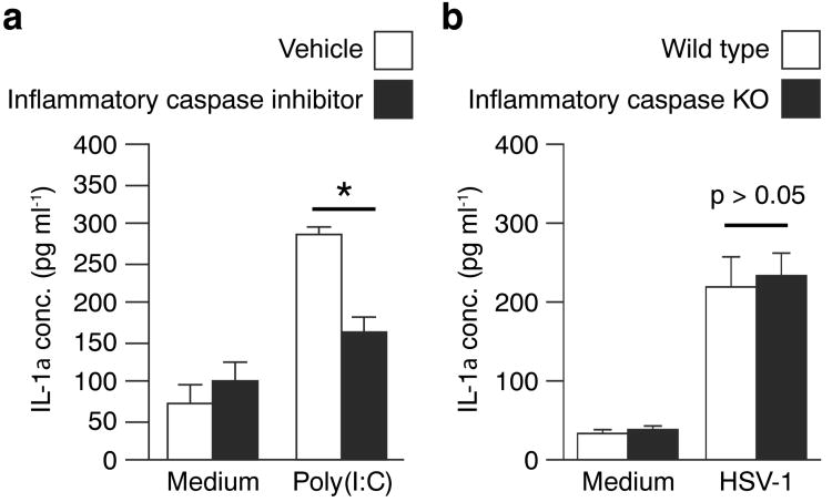
Figure 4. Poly(I:C), but not HSV-1, stimulated IL-1α release is inflammatory caspase dependent.
(a) Human primary keratinocytes were pre-treated with vehicle (white bars) or an inflammatory caspase specific inhibitor (black bars) for 30 min. before addition of medium or 25 μg ml-1 poly(I:C). Culture medium was collected after 12 hours and IL-1α levels determined by ELISA. (b) Mouse primary keratinocytes isolated from wild type (white bars) and inflammatory caspase deficient (black bars) mice were infected with HSV-1 (0.2 MOI) as described in Fig. 1. Control cells were treated with medium only. IL-1α released into the culture medium 24 hours post-infection was quantified using a mouse IL-1α specific ELISA. (a and b) Data points (n = 3) represent means ± s.d. Each experiment was repeated at least twice with similar outcomes. *, P < 0.05 (compared to medium only, t-test); **, P < 0.01.

