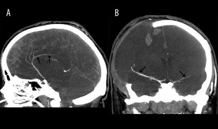
Figure 1.
The case of a 22-year-old woman with brain stem ischemic stroke and right-sided craniectomy presenting signs of BD on clinical examination; (A) – 10-mm MIP in the sagittal plane in CTA shows opacification of the right pericallosal artery (thin arrows); (B) – 10-mm MIP in the coronal plane in CTA shows opacification of M1 segments of MCAs (thin arrows).

