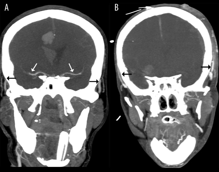
Figure 2.
Positive results of CTA in the diagnosis of BD: (A) – 10-mm MIP in the coronal plane shows stasis filling with delayed opacification of proximal MCAs (white arrows); please note the simultaneous opacification of the superficial temporal arteries (black arrows) (B) – 10-mm MIP in the coronal plane shows no intracranial filling; these findings confirm the diagnosis of BD.

