Figure 5.
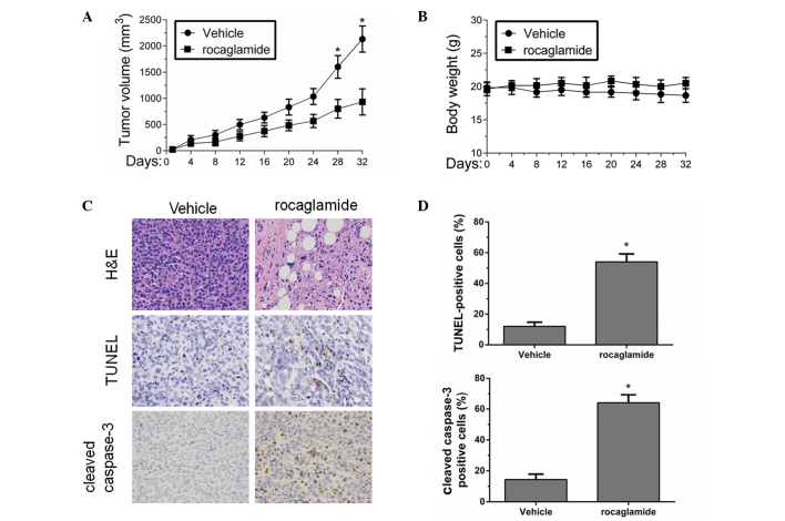
Effect of rocaglamide treatment on the growth of tumors from Huh-7 cells implanted in SCID mice. Huh-7 cell tumors established subcutaneously in SCID mice were treated with vehicle (5% DMSO in olive oil; n=5) or rocaglamide (2.5 mg/kg; n=5) via daily intraperitoneal injection into the mice for 32 days. (A) Tumor volume and (B) body weight were monitored. (C) Histological analysis of apoptosis in xenografted HCC tumors from the mice were performed using H&E, TUNEL and cleaved caspase-3 staining (magnification, ×200). (D) Quantitation of the TUNEL and cleaved caspase-3 staining in rocaglamide-treated tumors compared with vehicle-treated tumors. Data are expressed as the mean ± standard deviation. *P<0.01 compared with vehicle. Data are representative of three experiments. SCID, severe combined immunodeficient; DMSO, dimethyl sulfoxide; H&E, hematoxylin and eosin; TUNEL, terminal deoxynucleotidyltransferase-mediated dUTP nick end labeling.

