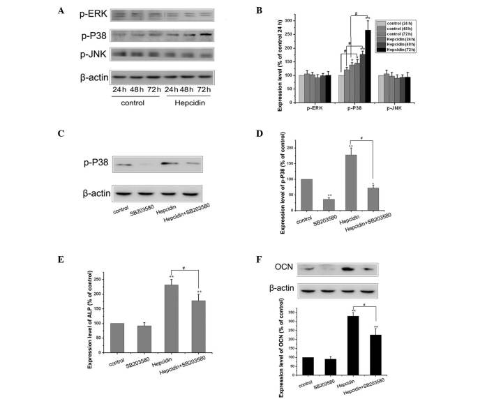
Figure 4.
MAPK/P38 signaling pathways contribute to osteogenic differentiation induced by hepcidin. (A) Cells in the control, siRNA control group and BMP2-siRNA groups were cultured in osteogenic medium with or without 0.2 mmol/l hepcidin for 48 h and the expression levels of p-ERK, p-P38 and p-JNK were detected using wetern blot analysis. (B) Each value is expressed as the mean ± SEM (n=3) and β-actin was used as a loading control (*P<0.05, vs. control; **P<0.01, vs. control; #P<0.01, vs. hepcidin. (C) Cells were cultured in osteogenic medium and exposed to 0.2 mmol/l hepcidin with or without 10 μM SB203580 for 48 h and the expression levels of p-P38 were detected by western blotting. (D) Each value is expressed as the mean ± SEM (n=3) and β-actin was used as a loading control (*P<0.05, vs. control; **P<0.01, vs. control; #P<0.01 hepcidin, vs. hepcidin + SB203580). (E) Cells were cultured, as described above, and the expression levels of ALP were detected using an alkaline phosphatase assay kit. Each value is expressed as the mean ± SEM (n=3; **P<0.01, vs. control; #P<0.01 hepcidin, vs. hepcidin + SB203580). (F) Cells were cultured, as described above, and the expression levels of OCN were detected by western blotting. Each value is expressed as the mean ± SEM (n=3) and β-actin was used as a loading control (**P<0.01, vs. control; #P<0.01 hepcidin, vs. hepcidin + SB203580). MSC, mesenchymal stem cell; MAPK, mitogen-activated protein kinase; RT-qPRC, reverse transcription polymerase chain reaction; SEM, standard error of the mean.

